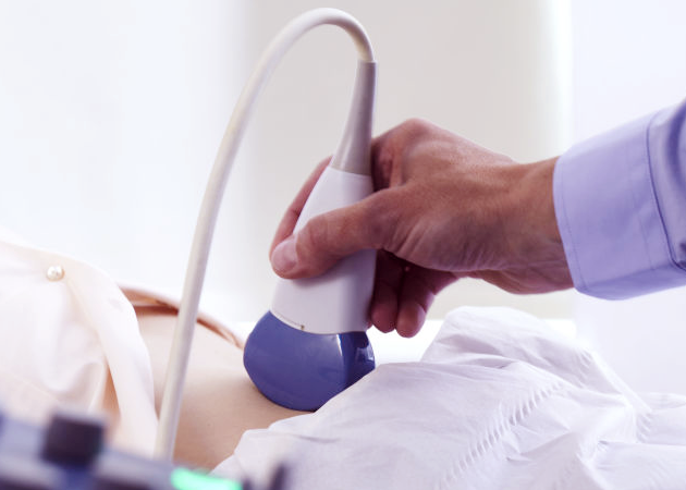
Ultrasound is a medical imaging technique that uses inaudible, high frequency sound waves to form images of the body’s internal organs and tissues. The sound waves are transmitted into your body using a small handheld transducer or probe, reflected off organs and other structures back to the probe, and converted into an image for display by a computer. Ultrasound images are displayed in real time, which means that the movement of organs, babies and even blood flow can be visualized and assessed.
Although ultrasound is most commonly associated with babies and pregnancies, it can be used to assess many structures in the body, some of which include (but are not limited to):
- Abdominal organs (liver, gallbladder, spleen, pancreas, stomach)
- Pelvic organs (kidneys, bladder, uterus, ovaries)
- Thyroid and parathyroid glands
- Scrotum (testicles)
- Breasts
- Arteries and veins
- Obstetrics (All stages of a developing pregnancy including Nuchal translucency genetic screening exam)
- Appendix, hernias and other lumps and bumps
- Heart
Most of the ultrasound exams above are also available for children, including the following pediatric only exams:
- Hip dislocation/dysplasia
- Cranium (head and brain)
- Spine
- Pyloric stenosis (stomach)
Types of Exams:
2D/3D/4D Real Time Exams
Technological advances in ultrasound allows us to image organs and structures in 2D, 3D or 4D (real time), which means that we can image a baby’s face in the womb and it is even possible to see your baby smile or yawn.
Doppler
An ultrasound technology that can be used to assess the blood flowing in your veins and arteries in order to detect blockages or clots. If your technologist uses Doppler, you will hear a gentle “whooshing” sound (like a heart beat) coming from the machine.
Interventional procedures
These include various types of biopsies, pain injections or fluid aspirations (drainage) where the needle placement is viewed in real time on an ultrasound machine to ensure accurate placement. In certain cases, biopsies may be available the same day to provide expedited results and optimal continuity of care for our patients
Echocardiogram
An echocardiogram (also called an Echo) is an ultrasound examination performed to assess the various components of your heart. The ultrasound images will show the size, shape, texture and movement of your heart muscles, the different heart valves, as well as the size of your heart chambers and how well they are working.

What to Expect
We make it a priority to ensure that you are at ease during your ultrasound exam.
- Complete the necessary medical forms obtained from front desk staff.
- Canada Diagnostic Centres provides private and secure change rooms (at most locations).
- Comfortable two piece scrubs will be provided for you to change into (at most locations). You may be asked to remove your jewelry.
- One of our friendly technologists will take you to the exam room and position you on a padded table.
- Warm, water based gel will be applied to your skin to help improve the quality of the pictures and a small handheld transducer or probe will be moved over the area of interest.